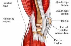
(see glossary for anatomical terms)
During your career as a cyclist, your knees may bend and straighten 162-324 million times (90 rpm x 60 min x 15-30 hrs x 40 wks x 50 yrs). One key to your cycling health would be maintaining a healthy knee.
The tools known as rollers or Sticks, promoted in endurance athlete-oriented catalogues, are suggested for knee maintenance. These tools, designed to compress the iliotibial band or tract (ITB) and adjacent tissues, create a more flexible structure. The restored flexibility/fluidity allows plasticity and relieves the tension–the mechanism theorized as creating symptoms. If anyone has experienced ITB syndrome, they may be acquainted with the more intense therapeutic devices known as tennis balls, baseballs or softballs (a misnomer, when used properly on the ITB).
But any tool’s effectiveness depends on HOW it is used. Athletes can roll their ITB and get little or optimal benefit. As an analogy, consider an expert carpenter and a child both using hammers. The carpenter can build a house in a week, whereas the child will simply bang on the board missing the nail for that same amount of time.
Most people rolling their ITB will be more like the child – they have the equipment and the motion, but are missing the nail. The nail, or target, in this case is a specific pressure threshold necessary to deform the tissue, which creates a piezoelectric property, and the subsequent release of pressure allowing for interstitial fluid re-entry. Both result in improved tissue. The intensity of pressure and its duration may be the most important element.
Today, you will become the expert carpenter. To be expert means understanding the material you use.
A quick review of the knee and ITB. The knee actually consists of two joints. The main joint associated as the knee is the tibiofemoral joint–the “hinge” of the femur and tibia bones. The other joint—patellofemoral–consists of the patella and the end of the femur.
A major contributor of force to the knee involves the ITB, which is located along the outside of your thigh. The ITB is not muscle but fascia, a thick collagenous/tendinous/ligamentous connective tissue. It’s strength and durability allow parts of it to be used for surgical repairs elsewhere in the body. Functionally, the ITB forms the tendon attaching lateral hip muscles to the length of the femur and at the knee.
The ITB can be a cyclist’s best friend. It stabilizes the lateral thigh, transfers force through its fibers, and counter-acts medial shearing associated with an overly strong vastus medialis. (the easily identifiable teardrop quadriceps muscle head prominently displayed on cyclists) This balancing act between these two dominant, strong forces maintain the patella in its groove — literally and metaphorically.
But the ITB can also be a cyclist’s worst enemy. Twenty-five percent of cycling soft tissue injuries originate from ITB Syndrome (ITBS) or ITB Friction Syndrome (ITBFS). As stated above, ITB tension is the proposed mechanism creating friction between the band and underlying structures. Symptoms can range from sore knees to a searing pain in the lateral side of the knee, so debilitative as to prevent putting pressure into the pedals or walking up and down stairs. The present surgical intervention alleviates the symptoms by removing the highly innervated fat pad underneath the band.
Fortunately, preventive and conservative therapeutic measures — such as using the roller and balls – can work to relieve tension, and thereby help minimize ITBS, and possibly even the need for surgery (though there are non-responders to conservative treatment).
Now, HOW to use a roller or ball on the ITB.
One of the best ways to learn movement is modeling—viewing someone learning a movement. So, instead of simply reading about proper technique for rolling your ITB, watch the following videos on YouTube.com.
These videos are for teaching purposes only. Their use implies no endorsement of the businesses, aesthetics, or philosophies associated with them. They are chosen simply because they are good demonstrations of the underlying principles.
Video 1
http://www.youtube.com/watch?v=3X0FOyXW7Vs&feature=related
As narrated in this video, the initial session and pressure response can be intense. Unfortunately, the required pressure threshold to deform tense tissue may be high, so high as to bring tears to one’s eyes.
Video 2
http://www.youtube.com/watch?v=AAuv9BUq_yY&feature=related
In this video, an intense, point-specific modality—a ball—demonstrates a more precise method. The pain increases as the ball concentrates the force, rather than spread out over a roller. This tool may be too advanced for acute symptoms.
If direct pressure creates intense pain, an athlete could start instead by attending to the surrounding muscle such as the vastus lateralis (the large quad muscle on the outer side of the thigh). This can be done with the roller or ball–as in the vastus intermedius/rectus femoris sections of the second video. These muscles could be adding indirect tension into the band. By addressing them, the athlete can ultimately reduce the tension in the band and then treat the band itself.
Video 3
http://www.youtube.com/watch?v=-GY0enibxJg
This video illustrates the importance of releasing associated muscles by addressing the posterior fibers of the gluteus medius. The only tool for this is the tennis ball.
Any type of proper rolling/massage included in an active lifestyle as an ongoing maintenance program prevents the ITB from reaching symptom-causing tension. Similar to stretching hamstrings, quads or psoas, rolling could be a daily addition to any effective cycling training program. BUT, if you are going to roll your ITB, remember that HOW you do it is what matters.
Scot Willingham, MA, ACSM, CSCS, USAC is a neuromuscular re-educator that coaches cyclists and other endurance athletes while working as a strength and conditioning rehabilitation expert in NYC. He is a recent graduate of Teachers College, Columbia University’s Motor Learning department. Motor Learning is the field that researches human motor control and is used as the foundation for many pedagogical decisions in physical education, sports coaching, movement teaching and rehabilitation.
THIS MATERIAL DOES NOT CONSTITUTE MEDICAL ADVICE. IT IS INTENDED FOR INFORMATIONAL PURPOSES ONLY. PLEASE CONSULT A PHYSICIAN FOR SPECIFIC TREATMENT RECOMMENDATIONS.
References:
Fairclough, J. et al. (2008). The functional anatomy of the iliotibial band during flexion and extension of the knee: Implications for understanding iliotibial band syndrome. Journal of Anatomy, 208, pp. 309-316.
Benjamin, M. (2009). The fascia of the limbs and back – a review. Journal of Anatomy, 214, pp.1-18.
Glossary and Tech Specifications
Anatomy illustration for visual reference:
This plate is mis-labeled as the medial (inner) side of the leg. It shows the outer side of the leg.
http://www.anatomyatlases.org/atlasofanatomy/plate14/02medlowerlimb.shtml
Condyles – the wider parts or platforms at the end of the femur and the top of the tibia that create the tibiofemoral joint (the hinge of the knee).
http://en.wikipedia.org/wiki/Lateral_condyle_of_femur
Distal – further away from the center.
Fascia – a global term describing many types of a collagenous connective tissue throughout the body; in this instance, the stronger, thicker fascia typically described as deep fascia.
Fascia lata – the fascial wrapping around the thigh.
http://en.wikipedia.org/wiki/Fascia_lata
Femur – thigh bone.
Gluteus group – the gluteus maximus, mediuus, & minimus (hip muscles).
Gluteus maximus – large hip muscle attaching at the sacrum to the femur and the iliotibial band.
Iliotibial band or tract – large band of thick fascia that originates from the gluteus group, the iliac crest (part of the pelvis) and the tensor fascia lata, covering and connected to the outer edge of the femur, and the fascia lata, attaching to the outer top of the tibia and the patellar retinaculum.
Intercondylar groove – groove on the front of the lower end of the femur that the patella (hopefully) glides over.
Ilium – the crested bone of the pelvis.
http://www.anatomyatlases.org/atlasofanatomy/plate03/02pelvisanterior.shtml
Lateral – away from the midline, i.e. in this article, the outer side of the thigh.
Patella – the knee-cap.
Patellar retinaculum – fascial connections and bundles around the patella.
Patellofemoral joint – the back of the patella and the intercondylar groove in the femur.
Rectus femoris – two-joint muscle of the quadriceps group.
Tibia – the “shin” bone of the lower leg.
Tensor fascia lata – internal hip rotator muscle on the anterior of the pelvis.
Tibiofemoral flexion – the angle of the hinge joint closing (bending the leg).
Tibiofemoral extension – the angle of the hinge joint opening (straightening the leg).
Tibiofemoral joint – the knee, the meeting of the femur and the tibia.
Vastus lateralis – outer muscle of the quadriceps (front of thigh).
Vastus intermedius – deep central muscle of the quadriceps.
Vastus medialis – inner muscle of the quadriceps.

hi alex, is me Antonio remember the guy from CRCA on monday in central park???….woaooo, very nice article, lots of information, thanks to write things like this….see u in the park during the week maybe at 10am ok….see ya…
best thing i’ve read on nyvc all year. incredibly helpful.
thank you for posting this.
Absolutely enlightening, thanks guys
i heard CCNS is going to becoming out with informational videos on different stretches for cyclists soon
you heard that, huh?
Cycling is considered as the one of the best exercise. During cycling all our muscles in our body will get stimulated. The iliotibial band or tract is helpful for the click this link people having pains on the joints in knees.
Thanks for sharing these videos, particularly the third one. Tennis balls can really be very useful. Leo of leosrackets.com